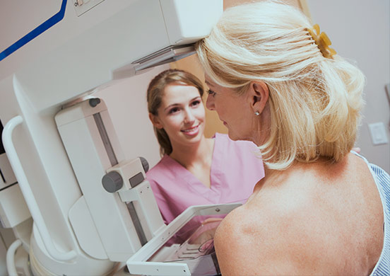
Mammography
Breast exam with the goal of detecting possible abnormalities using very low-dose x-rays.
Available at the
Maisonneuve and Bélanger clinics
About the Exam
A mammogram consists of x-raying each breast from the front and side to view the mammary gland in its entirety. Taking approximately 10 minutes, the mammogram enables the detection of such abnormalities as shadows or microcalcifications.
A mammogram does not always enable a definitive diagnosis; it shows whether or not an abnormality is present in the breast, but it does not enable a person to determine with certainty whether or not the abnormality is cancerous. Additional exams are then necessary to confirm a diagnosis:
- Special views (Magnification/Compression)
- Breast ultrasound
- Sample (biopsy)
Preparation

No preparation is necessary, however you must bring the images and reports from your last mammogram and/or ultrasound with you if it (they) was (were) performed in another centre. These previous records enable the radiologist to compare and note any changes that have occurred since the previous exam.
Do not apply antiperspirant or body cream the day of the exam.
Contraindication:
Within the context of a screening mammogram, before your appointment, if you observe irritation in the skin below the breast, we recommend that you postpone your appointment in order to let the skin fully heal.
Indeed, if your skin is irritated, there is a risk of tearing below the breast. Do not undergo this exam if you are pregnant.
Exam Process
When you arrive, we will ask you to fill out a questionnaire, which will be verified by our technologist.
For the exam, the technologist will place the first breast in the mammography unit. The breast is progressively compressed. Once the position of the breast is verified, an x-ray image is taken. You must be completely still and hold your breath at this time. The first breast is released and the operation is repeated on the second breast.
During a basic mammogram, if you have breast implants, 8 images will have to be taken instead of 4 images, because another technique is used in order to prevent the risk of rupturing the implant.
What You May Experience
This very quick exam can be uncomfortable due to the pressure put on the breast. This varies depending on each person’s sensitivity.
After the Exam
Additional exams are often required. However, you need not be alarmed as these exams are often performed to clarify the routine images. The results of your exam will be sent to your referring physician, who will take care of any necessary follow-up.
Other Services Offered
General Radiology
(Without an appointment)
X-ray medical imaging technique
Ultrasonography
Ultrasound-based medical imaging technique
Injection Therapy
Injection of an anti-inflammatory or lubricating product
Barium Swallow
Study of the upper digestive tract
Mammography
Study of mammary glands using x-rays
Osteodensitometry
Measurement of bone density
Barium Enema
X-ray exam of the large intestine
Small Bowel Follow-Through
Study of the lining of the small intestine
Cardiac ultrasound
Medical imaging test that uses ultrasound to study the anatomy and function of the heart and its structures
Contact us
Do you still have unanswered questions? Contact us. Our team will be happy to answer.
